Electron microscopy
The Central Electron Microscopy Facility provides state-of-the-art instrumentation, techniques, and expertise for advanced electron beam micro characterization of materials at high spatial resolution. Capabilities include microstructural and microchemical characteristics and materials properties; advanced electron microscopy techniques; computer analysis and instrumentation; and advanced education for students and investigators in the theory and application of electron optical techniques.
Electron and ion microscopy

- FEI Quanta 200 FEG SEM
A versatile, high-performance instrument with three modes (high vacuum, low vacuum, and ESEM) to accommodate the widest range of samples of any SEM system. It is optimized as a dedicated OIM (Orientation imaging by Electron Backscatter Diffraction) microscope. - FEI Quanta 600 FEG SEM
A versatile, high-performance instrument with three modes (high vacuum, low vacuum, and ESEM) to accommodate the widest range of samples of any SEM system. - Tescan Mira 3 FEG SEM
A field emission scanning electron microscope offering multiple imaging modes for high-resolution imaging, wide field-of-view, or large depth-of-focus. - 
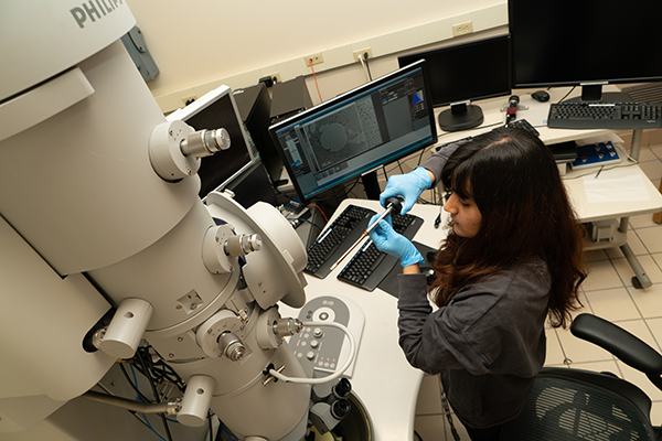
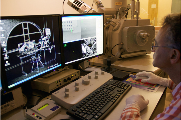
FEI Tecnai F20 TEM/STEM
Designed to produce optimum high-resolution performance in both TEM and STEM. It is optimized for materials applications that require either high-resolution STEM performance (imaging and spectroscopy by Electron Energy Loss Energy and Dispersive XRAY) or correlated imaging and analytical methods (for TEM and STEM). - FEI Nova Nanolab 600 Dual Beam FIB
- FEI Helios Xenon Plasma Dual Beam FIB
- Thermo Fisher Themis 200 G3 Aberration Correct STEM
The Themis 200 is a state of the art 80-200 kV FEG Scanning Transmission Electron Microscope designed to deliver ultimate performance in TEM and STEM modes - Thermofisher Themis TEM/STEM
Contact: Daniel Flaherty for more information

X-ray diffraction and tomography
The Materials Characterization Facility houses several x-ray devices including:
- Panalytical X’Pert Pro MPD X-Ray Diffractometer
The MPD uses focusing optics for analyzing powder and bulk materials. It has a PIXcel detector for rapid scanning. - Panalytical X’Pert Pro MRD X-Ray Diffractometer
The MRD uses lens, mirror and high resolution optics for parallel beam techniques. It is used for thin film analysis, pole figures, stress measurements, in-plane scans, reflectivity, rocking curves and reciprocal space maps. - Malvern Panalytical Empyrean XRD
The Empyrean has focusing optics and parallel beam optics. It is used for powder/bulk XRD analysis, thin film analysis, pole figures, residual stress measurements, reflectivity, rocking curves, microdiffraction, and in situ high temperature work.
- Additional X-Ray Equipment
The Materials Science and Engineering Department also maintains x-ray instruments in laboratories outside the central facility.
- Xenocs Xeuss 3.0 SAXS/WAXS
This instrument supports the development of cutting-edge nanostructured materials that have keys features ranging in size from tens of Angstroms to hundreds of nanometers. To allow in situ and in operando characterization of structural properties, this custom, automated Xenocs Xeuss 3.0 x-ray scattering system enables high-throughput studies at a variety of length scales.
- Xenocs Xeuss 3.0 SAXS/WAXS
-
- 3D Computed Tomography
Contact: Betsy Clark for more information
Scanning probe microscopy

NT-MDT Solver NEXT SPM
This is a general purpose SPM that is capable of preforming AFM and STM. With the convenience of an all-automated setup and control.
Download the NT-MDT Solver Next SPM Manual
NT-MDT NTEGRA SPM
This microscope has the same AFM capabilities as the Solver NEXT and more. Allowing the analysis of mechanical measurements, larger samples, samples immersed in liquid and a high-resolution scanner.
Download the NT-MDT NTegra SPM
NT-MDT Spectra AFM/Raman
The Spectra has similar AFM capabilities as the Solver and NTEGRA but with the added befit of a confocal Raman spectrometer. The Raman system is outfitted with a 532nm and 633nm laser paired with 4 different gratings. The combined AFM Raman setup allows for tip enhanced Raman (TERS), this allows for an improvement in spatial resolution from hundreds of nanometers to tens of nanometers.
Download the NT-MDT Spectra AFM/Raman
Contact: Betsy Clark for more information.
Optical Microscopy
Nikon Eclipse ME600
Zeiss Imager Z2 Vario
Zeiss Observer Z1m Inverted Microscope
Contact: Betsy Clark for more information.
Sample preparation

The Materials Characterization Facility includes a complete specimen preparation facility for transmission and surface specimen preparation. Supporting the specimen preparation for transmission electron microscopy are three Gatan Precision Ion Polishing machines, four dimpling machines, and an electro-polishing system. Surface preparation for scanning electron microscopy specimens includes high vacuum sputter coating and ion etching equipment. For advanced sample preparation, the The NovaLab 600 Dual Beam, Focused Ion Beam and Field Emission Scanning Electron Microscope, is available.
Contact: Betsy Clark for more information
Allied Multiprep Polishing System – 8”
- Precise semiautomatic polishing for a wide range of materials for microscopic (optical, SEM, TEM) imaging
- Capability for parallel polishing, angle polishing, or site-specific polishing
- Dual micrometers enabling quantifiable material removal monitored in real time
- Variable rotation and oscillation speeds
- Adjustable load control
Fischione Model 110 Automatic Twin-Jet Electropolisher
- Electrolytic polishing or chemical etching
- Simultaneous two-sided polishing
- No induced artifacts
- Adjustable flow rate, jet and specimen positions
Buehler EcoMet 250 Grinder-Polisher
- Manual polishing for a wide range of materials
- Wide array of polishing mediums available
- 8” platen size, 10-500 rpm speed, clockwise or counterclockwise
Buehler IsoMet 1000 Precision Saw
- Precision sectioning saw for cutting a variety of materials with minimal deformation
- Gravity fed cutting force
- RPM ranges from 100-975
- Table saw attachment for precision sample alignment
Gatan Precision Ion Polisher (PIPS) II
- X,Y stage permits alignment of argon beam to region of interest on the sample
- Argon ion milling with voltages between 0.1 to 8kV
- Liquid nitrogen cooling available
- Variable ion gun angles ±10°
- Touch screen for display and control of parameters
- Digital optical image with image storing and analysis in DigitalMicrograph software
Fischione Model 1020 Plasma Cleaner
- Cleans samples with high-frequency Argon/Oxygen plasma
- Simultaneously cleans TEM and SEM specimens and specimen holders
- Removed existing carbonaceous contaminations and prevents further contamination
- No material etching or sputtering
EMS Q300T D Plus Dual Target Sequential Sputtering System
- Suitable of sequential sputtering of two materials
- Fully automated with user defined recipes
- User controlled pumping sequence, number of sputter cycles, time, and current
- Adjustable stage allowing for various sample sizes and geometries
- Film thickness monitor for precise thickness control
- Interchangeable sputtering targets including gold, platinum, carbon, chromium, nickel, etc