
Improved muscle mapping could aid neurological treatment
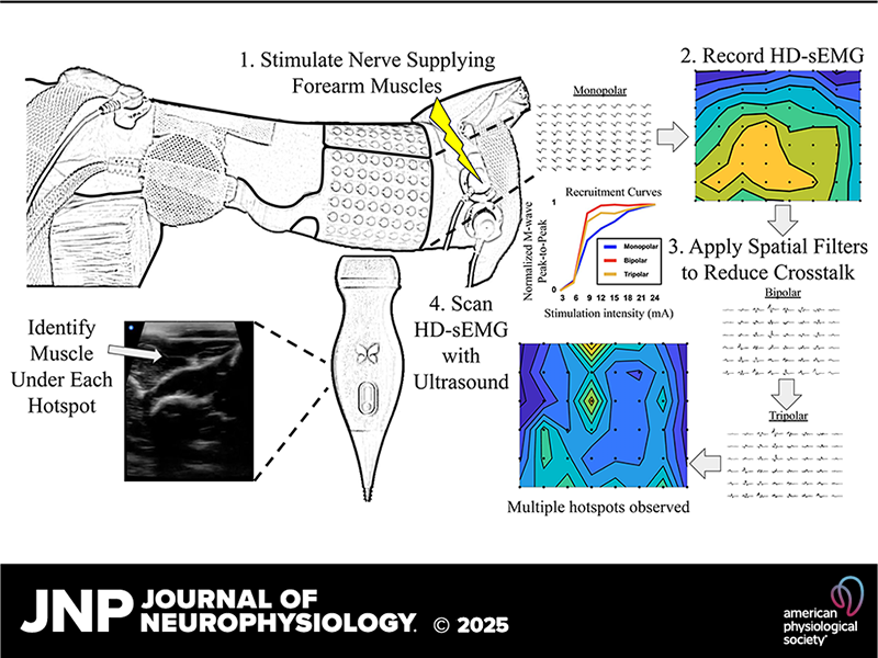
Carnegie Mellon researchers are using high-density surface electromyography (HD-sEMG) with spatial filters and ultrasound to reduce crosstalk and enhance muscle activity localization. This approach aims to achieve precise mapping of muscle activity, particularly in complex structures like the forearm.
Researchers from Carnegie Mellon University have developed a cutting-edge method to identify muscle activity in densely packed regions like the forearm. Using high-density surface electromyography (HD-sEMG) sensors alongside other techniques such as peripheral nerve stimulation, spatial filtering, and ultrasound imaging, this approach offers more accurate identification of muscle activity. The findings could lead to better treatments for neurological injuries and advancements in prosthetic limb control.
Source: Journal of Neurophysiology
By electrically stimulating specific nerves, researchers can selectively activate muscles, providing a controlled way to study muscle activity. The HD-sEMG system used in this study features a 64-channel grid that is adhesively applied to the skin to capture electrical signals, known as M-waves, produced by active muscle contractions. The sensors provide high-resolution measurements of muscle activity, allowing researchers to apply advanced spatial filters to minimize electrical interference from neighboring muscles, known as crosstalk, and to isolate M-waves from target muscles. Applying these filters to HD-sEMG nearly eliminated crosstalk at distances of 2.55 cm or more. Reducing crosstalk allows for clearer separation of hotspots on heat maps, making it easier for researchers to distinguish muscle activity and use ultrasound imaging to verify the location and identity of the underlying muscles.
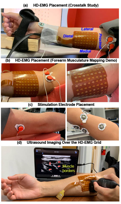
The HD-sEMG system used in this study features a 64-channel grid that is adhesively applied to the skin to capture electrical signals, known as M-waves, produced by active muscle contractions.
Accurately identifying the strength and location of muscle activity with minimal distortion is critical for studying motor function, especially for diagnosing problems caused by stroke, spinal cord injury, and other neurological disorders. The techniques developed in this research have the potential to improve neurological treatments, such as physical rehabilitation, as well as improve the control of prosthetic limbs.
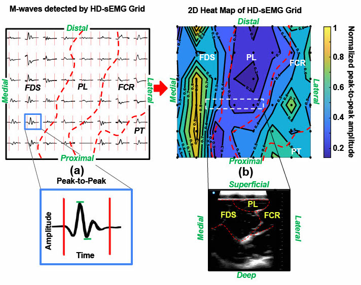
Reducing crosstalk allows for clearer separation of hotspots on muscle activity maps, making it easier for researchers to distinguish different sources of muscle activity and use ultrasound imaging to verify the location and identity of the underlying muscles.
“We are currently applying this method to clinical populations, including stroke patients with hemiplegia and amputees with phantom limb pain,” explained Ernesto Bedoy, a postdoctoral researcher at CMU who led this study. “We’re using this approach to better understand muscle activity patterns in these populations and develop personalized treatment strategies that maximize recovery.”
This research was published in the Journal of Neurophysiology. The researchers include Bedoy; Douglas Weber, a professor of mechanical engineering and member of the Neuroscience Institute; and Efrain Guirola Diaz, Ashley Dalrymple, Isaiah Levy, Thomas Hyatt, Darcy Griffin, and George Wittenberg.