A handheld device to monitor breast cancer lesions
Biomedical engineering Ph.D. student Constance Robbins is creating a breast lesion monitoring device that has the potential to revolutionize breast cancer treatment worldwide.
Breast cancer is the most prevalent non-skin cancer among women. Starting as an abnormality within the breast, the cancer is at risk of spreading to the rest of the body; therefore, early detection of such abnormalities is paramount. Equally as important as detecting them is monitoring and studying them regularly for changes—but do we have the tools necessary to do this?
The answer is yes, and no. As of now, most of the advocated procedures for both diagnosis and monitoring—mammograms, breast ultrasounds, breast biopsies, breast MRIs—require highly expensive equipment. It’s not feasible for these procedures to be performed regularly, or in underdeveloped areas where access to such equipment is rare. And although breast self-examinations, or BSE—wherein one touches the breast to detect lumps—is a highly advocated, inexpensive way to first detect lesions, it does not provide information that can be recorded and tracked.

Source: Constance Robbins
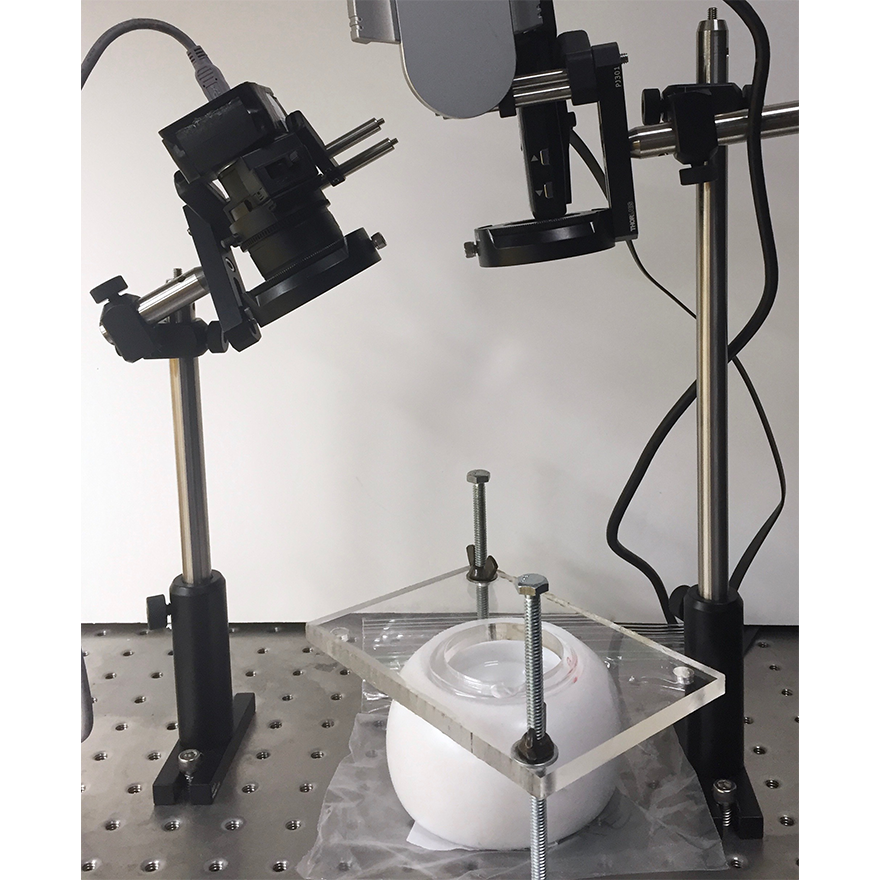
Model of imaging and compression system that Robbins demonstrated at SPIE conference. Here, a petri dish is placed underneath an acrylic plate. In the top left, a CCD camera is mounted and pointed at the petri dish. In the top right, a laser projector also points down.
Thus was born the need for an alternative method—and responding precisely to that need is second-year Ph.D. student Constance Robbins, who is working with Jana Kainerstorfer, assistant professor of biomedical engineering, to develop a new device. James Antaki, professor of biomedical engineering, is also a collaborator on this project.
“We’re not trying to replace mammograms,” says Robbins. “If you have results that are inconclusive, but there’s not high enough of a risk to justify a biopsy right away, then that would be something we could use this device for.”
The first iteration was called “Palpaid.” A project developed by Molly Blank, a former biomedical engineering Ph.D. student advised by Antaki, Palpaid was intended to be a small handheld device that measured the mechanical properties of a lesion. This iteration of the device relied on the relative stiffness of the lesion compared to its surrounding tissue. When pressed against the breast, the device’s flexible, reflective lens would deform around the stiff tissue and capture a topographic image of the deformation, which could then be examined for information on size, shape, stiffness, and location.
We’re not trying to replace mammograms. If you have results that are inconclusive, that would be something we could use this device for.
Constance Robbins, Ph.D. student, Biomedical Engineering, Carnegie Mellon University
Robbins decided to take Palpaid one step further, hoping to measure not only mechanical properties but vascularization as well. Breast lesions are stiff and highly vascularized, meaning that they contain greater total hemoglobin concentration than the surrounding softer tissue. But how can something like blood content be measured non-invasively?
The answer is with light. Robbins is using an imaging method called spatial frequency domain imaging (SFDI), where different patterns of light are shown on an area to extract information from the way those patterns are reflected differently. The variations in reflection tell us the absorption coefficient of the tissue, or how much light the tissue can absorb, which in turn depends on the concentration of hemoglobin in the tissue. However, light can only penetrate so far past the surface of the breast—what if a lesion is too deeply embedded?
“We’re trying to measure the absorption coefficient of the stiff tissue as well as the healthy tissue around it,” says Robbins. “But if the lesion is too deep, we won’t be able to detect it—that’s why we use compression, to decrease the distance from the lesion to our sensors.”
By compressing the tissue and essentially pushing the lesion momentarily closer towards the surface of the breast, the light can penetrate further into the tissue, and the device can get a better read on the lesion’s blood content.
Robbins presented her research at the SPIE Photonics West Conference, a conference dedicated to optics, in January of this year. Using a Tupperware container as a mold to create a slab of silicone that mimicked a breast, Robbins embedded multiple stiff inclusions within the fake breast at different depths. She demonstrated that by using compression, the inclusions became much more visible than when uncompressed.
At the conference, Robbins also encountered some constructive skepticism regarding her project, where people were unsure that SFDI could be applied to such a task. But she hasn’t lost faith. As a second-year Ph.D. student, she’ll be finished with her required coursework by next year and will be able to devote her time entirely towards research. Having already started the next round of experiments, Robbins hopes to effectively create a consumer electronic device that has the potential to revolutionize breast cancer treatment worldwide.
