Bioelectric Medicine Initiative
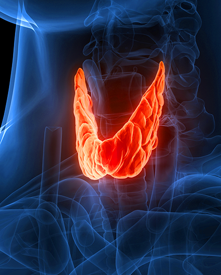
Researchers aim to develop a swallowable smart pill that temporarily operates in the gut to gently adjust nerve activity linked to stress.
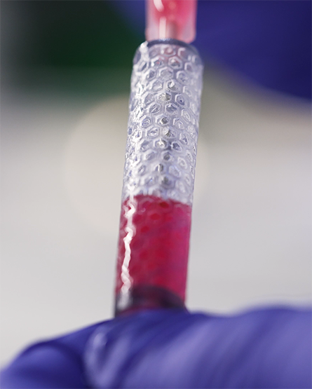
A multi-institution team has developed an effective intervention to shield a sufficient number of cells from the host immune system, while also providing access to oxygen and nutrients.
A new study shows ultrasound can subtly prepare the brain to respond, rather than directly triggering activity. Combined with light electrical stimulation, it produces stronger, targeted effects for future therapies.
A machine-learning method analyzes all major epilepsy biomarkers noninvasively, offering a faster, unified way to locate seizure-origin regions.
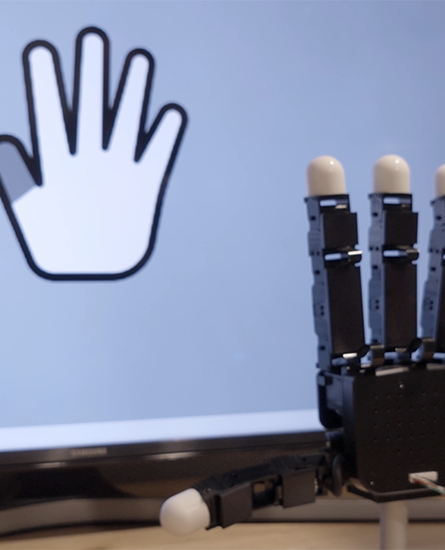
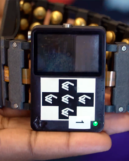
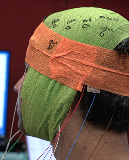
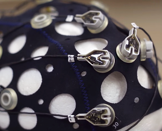
New research explores the transformative potential of EEG-based BCIs and their application beyond basic communication to intricate motor control.
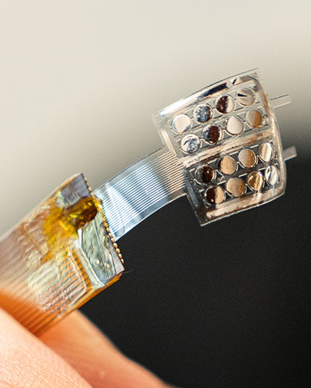
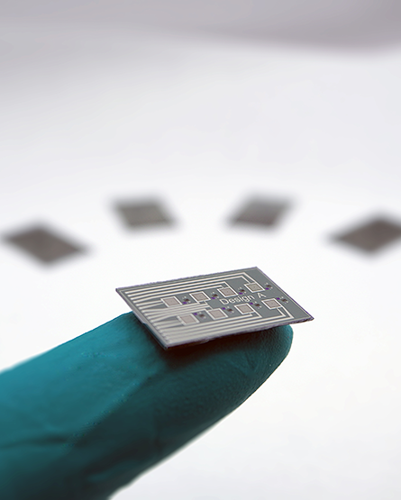
Wound assessment can be challenging due to its subjective nature, but a new sensor array quantifies biomarkers and has potential to offer real time measurements that could improve the healing process.
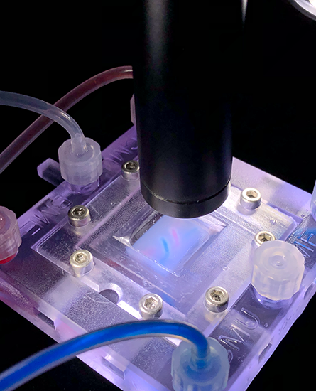
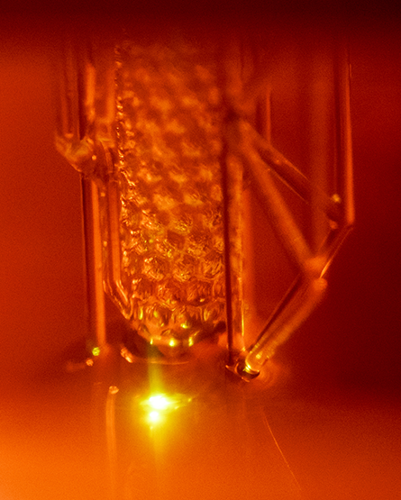
Using their novel FRESH 3D bioprinting technique, which allows for printing of soft living cells and tissues, the Feinberg lab has built a first-of-its-kind tissue model entirely out of collagen.
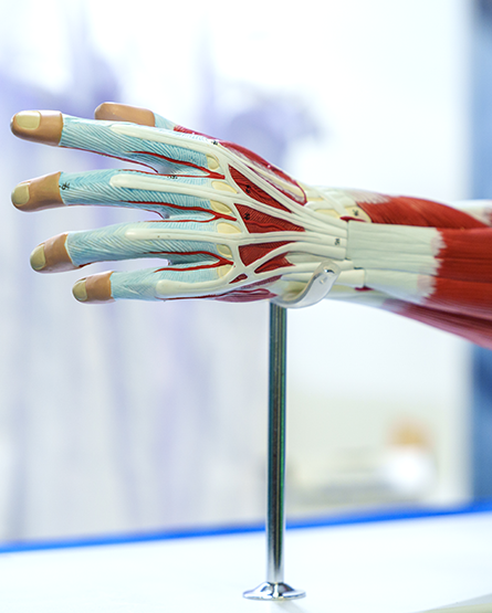
Carnegie Mellon researchers are using high-density surface electromyography (HD-sEMG) with spatial filters and ultrasound to reduce crosstalk and enhance muscle activity localization. This approach aims to achieve precise mapping of muscle activity, particularly in complex structures like the forearm.
Marc Dandin receives prestigious five-year grant given to junior faculty for research and education.
Carnegie Mellon University researchers have developed a new method for deep brain stimulation called “DeepFocus” that is minimally invasive and could treat conditions like PTSD and addiction.
Center for Neural Basis of Cognition researchers designed a clever BCI experiment to determine whether one-way activity paths, long hypothesized by neural network models, are used in the brain.
Keith Cook joins a multi-institutional, DARPA-funded project to create a novel ECMO and advanced life-support system device capable of rapid deployment to support wounded military personnel.
A CMU-led project team secured an award of up to $42M from ARPA-H to accelerate the development of implantable bioelectronic devices that deliver patient-specific therapy and monitor disease status.
A CMU-led team of researchers has secured an award of up to $34.9 million from ARPA-H to develop a new bioelectric medicine-based treatment for obesity and Type 2 diabetes patients.
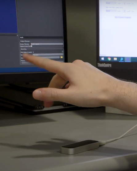
In collaboration with Meta, Doug Weber’s lab is exploring how sEMG signals can enable people with spinal cord injuries to interact with computer and mixed reality systems.
To advance cell-based therapies, researchers have identified a novel device that makes on-site oxygen for biological cells transplanted inside the body.
Researchers from Carnegie Mellon University, the University of Pittsburgh, and the University of Cincinnati have combined their expertise in engineering and medicine to create a noninvasive method for detecting worsening brain injuries before they happen. This advancement could reshape neurocritical care.
ARPA-H has awarded $45 million to a multi-institutional team of researchers to rapidly develop sense-and-respond implant technology that could slash U.S. cancer-related deaths.
Spinal cord stimulation technology developed by Douglas Weber in collaboration with the University of Pittsburgh offers new hope for people living with impairments that would otherwise be considered permanent.
Intracranial pressure sensing is the burgeoning focus of Jana Kainerstorfer’s biomedical optics lab, and her team is working to create noninvasive ICP sensing alternatives.
Doug Weber and an international team of researchers detected electrical signals in paralyzed muscles, which could be used to control robotic assistive devices.
Using light to control how cells “talk” to one another isn’t new science, but researchers at CMU have discovered that MXene, an easily produced nanomaterial, can allow for effiicient cellular communication.
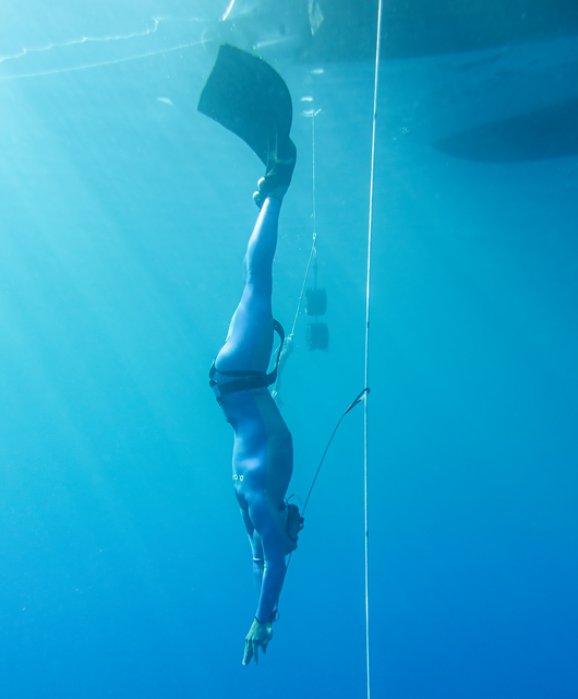
Carnegie Mellon University researchers are part of an international team working on wearable biomedical technology that will enhance freediver safety, as well as provide fresh treatment insights for cardiac patients.
Carnegie Mellon researchers are working with DARPA, Northwestern University, and Rice University to develop a system for regulating the body’s circadian clock.
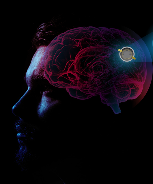
The team is creating a smart port to the brain that will use artificial intelligence to selectively stimulate and record from the brain.
Maysam Chamanzar’s team has developed a new class of materials for optical biointerfaces.
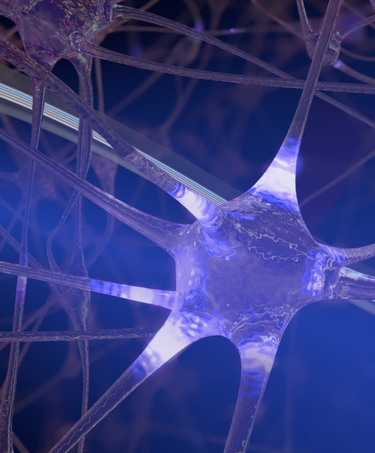
A novel material for controlling human neuron cells could deepen our understanding of cell interactions and enable new therapies in medicine.
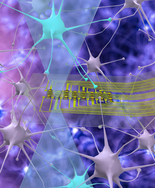
An interdisciplinary collaboration between researchers at CMU has produced a new type of neural probe with an innovative design, improving the way researchers study neurons deep in the brain.
One day, scopes may no longer need to be inserted into the body, such as down the throat or under the skin, to reach the stomach, brain, or any other organs for examination.
A team of researchers from Carnegie Mellon is starting a project to design and implement a high-resolution, noninvasive neural interface that can be used as a wearable device.
A team led by Pulkit Grover created more efficient deep neural networks called PolyDot coding to reduce errors and increase processing speed.
More than 50 million people of all ages suffer from epilepsy, the fourth most common neurological disease in the world.
Writers and scientists throughout history have searched for an apt technological analogy for the human brain, often comparing it to a computer.