A FRESH take on bioprinting
Olivia Olshevski
Apr 1, 2019
In the past five years, significant advances have been made in engineering biological tissue using 3-D bioprinting. Most recently, this method has been used to build tissues for applications in drug discovery and disease modeling, as well as tissue regeneration. However, despite the remarkable progress made in the application of bioprinting, the process remains technically challenging.
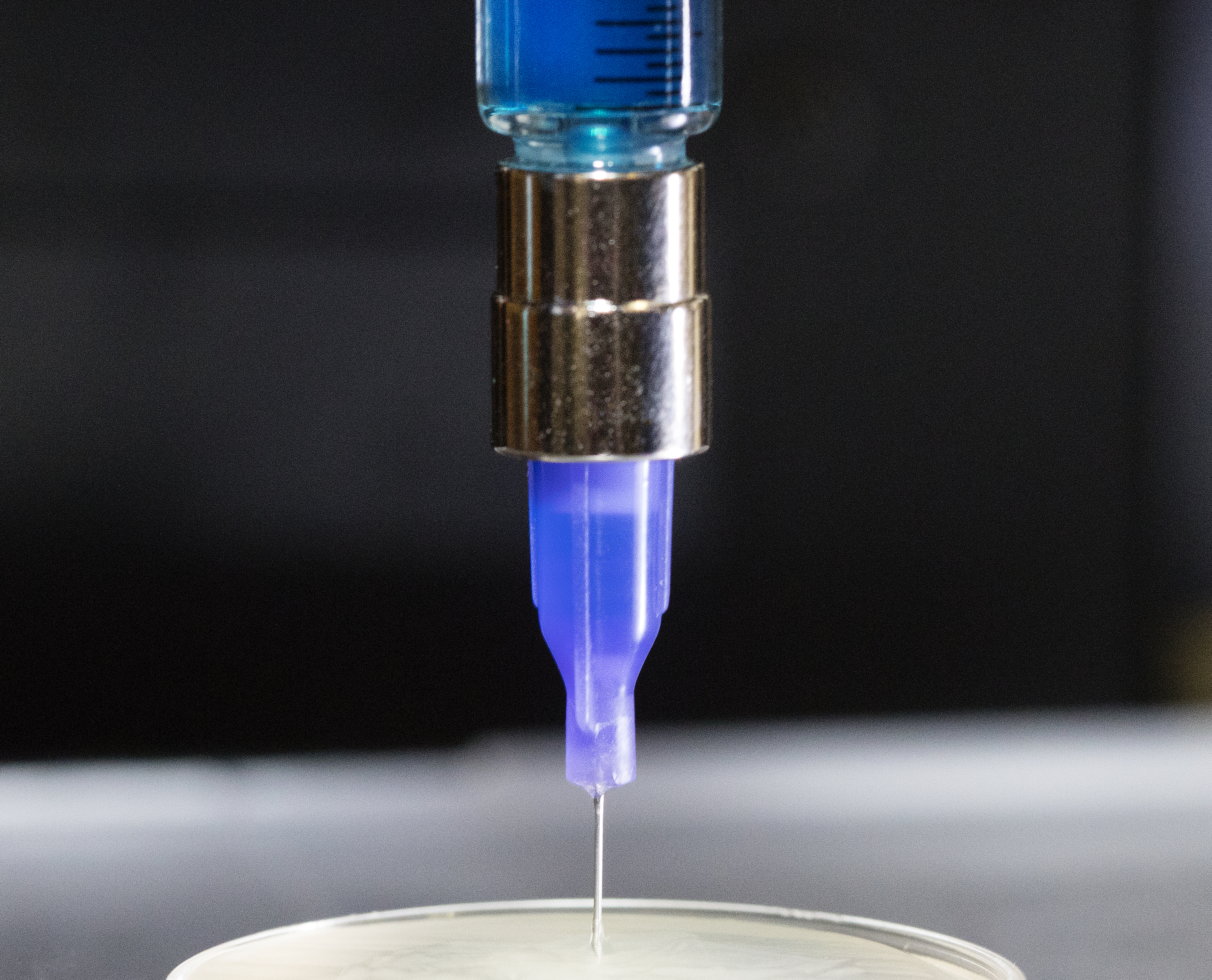
Though bioprinting is improving, poor fidelity and reproducibility are still major concerns. Recently, Adam Feinberg, an associate professor of biomedical engineering and materials science & engineering, has developed the novel Freeform Reversible Embedding of Suspended Hydrogels (FRESH) 3-D bioprinting technique, which allows for the printing of soft gels. These soft gels are printed inside of another soft support gel, ensuring that the printed structure does not collapse during the printing process. While this method is revolutionary in printing cellularized tissue scaffolds, defects often cannot be found until after the print has completed, wasting valuable cells and materials.

Source: Carnegie Mellon University's College of Engineering
To address this issue, Feinberg and Jana Kainerstorfer, an assistant professor of biomedical engineering, have joined forces to create a technique that allows users to monitor 3-D printed parts during the actual printing process.
Because the support gel is not fully transparent, it is difficult to detect printing errors by eye alone. Instead, Kainerstorfer has brought her expertise in optical imaging, using optical coherence tomography (OCT) to image the print throughout the entire process. Developed specifically for biological tissue, OCT is capable of visualizing through translucent tissue. By using OCT to monitor the prints, tissue engineers can optimize cost: OCT allows errors to be realized earlier in the process, meaning that less material is wasted. Additionally, Kainerstorfer hopes to adapt OCT and other optical methods to assess functional cellularization of these printed scaffolds and provide visual proof of viability.
By combining FRESH 3-D bioprinting and OCT, the researchers hope to produce a method of engineering biological tissue that enables researchers to monitor the printed part throughout the entire process and improve functional performance.
In the future, Kainerstorfer and Feinberg plan to add machine learning to the technique, which will enable users to predict when and where a print may fail during printing and even take corrective action to fix it in real time. This project is an important step in advancing 3-D bioprinting technology to the level already established in other 3-D printing fields.
