New equipment accelerates imaging process
Monica Cooney
Mar 20, 2024
In December 2023, a new high performance Raman microscope, the Horiba LabRAM Soleil, was added to the Materials Characterization Facility (MCF). This instrument is capable of ultra-fast imaging, up to 100 times faster than a conventional Raman spectrometer, and analysis automation. The microscope is equipped with four lasers and an ultra-low frequency filter to suit a wide variety of Raman spectroscopy needs.
Though the LabRAM Soleil is relatively compact, it offers one of the largest class 1 sample compartments available in microscopy tools. With this microscope, the MCF possesses Horiba’s full collegial support program. This allows users to contact Horiba’s in-house experts to address experiment specific questions.
“The capabilities of this instrument are difficult to rival,” explains MCF specialist Andrew Nickischer. “It can push the modern limits of Raman scattered light detection, all while being straightforward for users to learn and understand.”
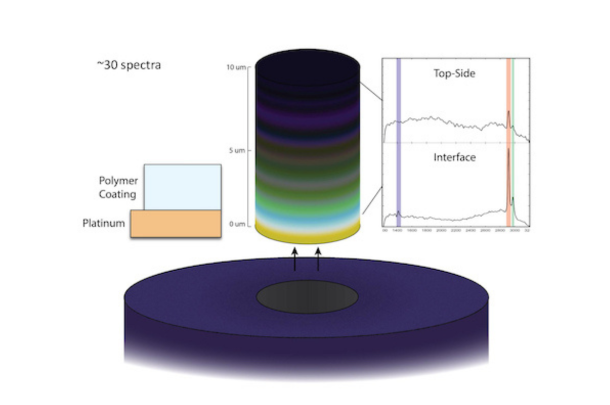
Source: Liyang Wang and Daniel Ranke
Sub-surface interfaces can be directly examined with high resolution through depth-resolved laser scanning. With this, differences in bonding states can be directly compared between bulk and interface Raman spectra to better elucidate interface organization. This is demonstrated here on an electrode stack of Pt and polymeric coating applied for sensing.
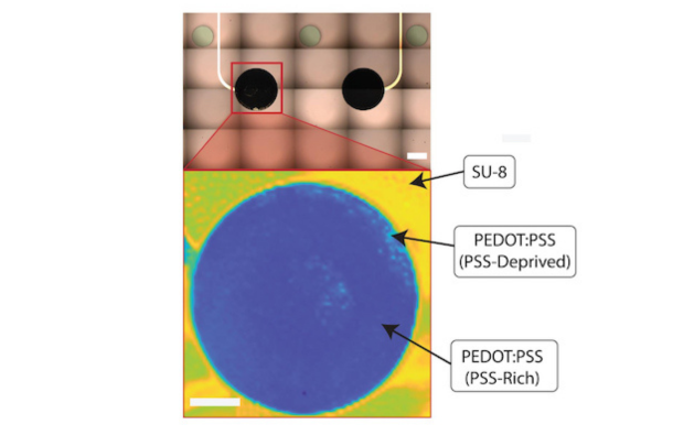
Source: Sam Gershanok and Daniel Ranke
Electrodes electrodeposited with PEDOT:PSS are imaged through >5600 high-quality Raman spectra to generate chemical maps that can be directly compared with optical imaging for quick and informative analysis of large-scale processes. (scale bars 250 and 125 um for optical and Raman map respectively). ~5600 spectra
“Through the Horiba Soleil, our group can quickly acquire high-quality three-dimensional chemical maps for analysis of electrodes for bioelectronics, chemical sensors, new hybrid nanomaterials, and more with a level of detail, scale, and informatics previously impossible,” said doctoral student Daniel Ranke, member of the Cohen-Karni research group.
MCF staff members have been trained on use of the Horiba LabRAM Soleil, and will be working to assist faculty and student users as they begin to utilize this tool in their research.